Swift Relief: Expert Treatment for Kidney Stones
If you are experiencing the sudden, severe pain of kidney stones, timely and effective care is critical. Northwoods Urology offers rapid diagnostic services and advanced treatments, including innovative laser lithotripsy, to quickly break up and remove stones. We are here to provide immediate relief and long-term prevention strategies.

Your Fast Track to Kidney Stone Treatment
We treat kidney stones with urgency and expertise. Our streamlined process ensures you receive effective diagnosis and treatment as quickly as possible to alleviate pain.
Understanding Kidney Stone Formation and Symptoms
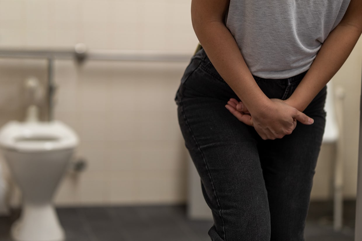
Kidney stones are hard deposits made of minerals and salts that form inside your kidneys. They are not always painful until they begin to travel down the urinary tract, which is when they can cause severe, debilitating pain.
Common Symptoms of Kidney Stones
- Severe Pain: Sharp, intense pain in the side and back, below the ribs, which often radiates to the lower abdomen and groin.
- Pain Fluctuations: Pain that comes in waves and varies in intensity.
- Urinary Changes: Persistent need to urinate, painful urination, or passing only small amounts of urine.
- Other Signs: Pink, red, or brown urine (blood in urine), and nausea or vomiting.
What Causes Kidney Stones?
Stones form when your urine contains more crystal-forming substances (like calcium, oxalate, and uric acid) than the fluid in your urine can dilute. Factors that increase your risk include family history, diet, dehydration, and certain medical conditions.
Advanced, Customized Kidney Stone Removal
We offer a comprehensive range of stone treatment options, from non-invasive methods to advanced surgical techniques, ensuring we can treat stones of any size and location with maximum effectiveness and minimal downtime.
Medication and Observation
For small stones (usually less than 4mm), we may recommend observation and medication to help the stone pass naturally. These medications, known as alpha blockers, help relax the muscles in your ureter, allowing the stone to pass more easily and with less discomfort.
Innovative Procedures
Our commitment to leading-edge technology means swift and precise stone removal.
- Laser Lithotripsy: A minimally invasive technique using a Holmium:YAG laser to vaporize all types of kidney stones, regardless of composition. It is one of the safest and most versatile intracorporeal techniques available.
- Extracorporeal Shock Wave Lithotripsy (ESWL): Uses sound waves from outside the body to break the stones into tiny pieces that can then be passed in the urine.
- Ureteroscopy: A tiny telescope is passed through the bladder and ureter to either remove the stone whole or break it up with a laser.
Real Patients. Real Stories. Real Results.
Read their stories to see how our personalized care and innovative treatments have helped people like you find relief and get back to living life to the fullest.


Frequently Asked Questions about Kidney Stones
When you are in pain, you need answers immediately. Review our frequently asked questions about kidney stones to learn more about our urgent care process and advanced treatment technologies.